
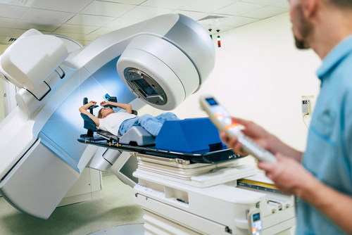
A study in the International Journal of Radiation Oncology – Biology – Physics evaluated predictors of liver segmental hypertrophy following radiotherapy for metastatic liver cancer.
The study analyzed data and imaging from 148 patients who underwent radiotherapy for primary metastatic liver cancer. Radiotherapy planning, baseline computed tomography (CT), and 3-month follow-up CTs were included. The researchers trained an nnU-Net-based model on the CT images to contour the liver segments (1, 2, 3, 4, 5-8). Ultimately, 52 features were collected, corresponding to segments 1, 2, 3, 2+3, 4, and 5-8, including equivalent dose to 2Gy fractions metrics-mean dose (Dmean), dose received by 95% of the volume (D95), volume spared from x Gy (Vx), cancer type, tumor location, and induction chemotherapy status. Six response variables were compared with potential predictors of hypertrophy using the Chi-squared/Fisher exact test (CST/FET) and logistic regression.
Hypertrophy by Liver Segment
The authors reported that segments 1, 4, and 5-8 demonstrated hypertrophy in 35% of cases. Segments 2, 3, and 2+3 demonstrated hypertrophy in 45%-49% of cases.
When stratified according to tumor location, segment 2+3 hypertrophy occurred in 66% of patients when the tumor was located in segments 5-8. In bilobed tumors, 34% of patients had segment 2+3 hypertrophy.
CST/FET analysis found that tumor location, induction chemotherapy, and tumor type were significant predictors of hypertrophy in segments 5-8. Tumor location was also a significant predictor for segments 3 and 2+3 hypertrophy.
Dosing and Hypertrophy
Logistic regression analysis found that all segment-based dose metrics significantly predicted segment hypertrophy, with the exception of Dmean in segment 4 and D95 in segments 2 and 4. The strongest predictive association was found for V35, which was significantly predictive of hypertrophy in each segment.
The mean dose for segments demonstrating hypertrophy was significantly lower, ranging from 15Gy to 30Gy, compared with segments with atrophy (P<.01); however, in segment 4 the mean dose was 10Gy lower than in segments with atrophy, a number that did not reach statistical significance. The authors noted that induction chemotherapy impacted the threshold mean dose that was associated with hypertrophy, “with more toxic drugs reducing the mean dose threshold.”
Overall, “tumor location and induction chemotherapy significantly impact the response of segments to radiotherapy. Dose volume metrics are strong predictors of volumetric response, with segment volume spared from 35Gy being the strongest predictor,” the authors concluded.


 © 2025 Mashup Media, LLC, a Formedics Property. All Rights Reserved.