
A new ultrasound technology that was recently tested could greatly improve the diagnosis of congenital heart disease in children and infants. The technique, known as vector flow imaging, was utilized by biomedical engineers from the University of Arkansas in a study published in a March edition of Progress in Pediatric Cardiology.
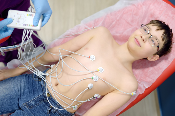
These researchers collaborated with cardiologists at Arkansas Children’s Hospital in Little Rock to test vector flow imaging in pediatric patients for the first time. This technique was used to generate comprehensive images of babies’ internal heart structures and blood flow. These images can be taken from any angle, and can be in still or dynamic format.
“Vector flow imaging technology is not yet possible in adults, but we have demonstrated that it is feasible in pediatric patients,” said Morten Jensen, associate professor of biomedical engineering at the University of Arkansas. “Our group demonstrated that this commercially available technology can be used as a bedside imaging method, providing advanced detail of blood flow patterns within cardiac chambers, across valves and in the great arteries.”
Alongside Jensen in the study was Dr. Hanna Jensen, a fellow University of Arkansas biomedical engineering professor, Dr. Thomas Collins, a pediatric cardiology professor at Stanford University School of Medicine, and other researchers from the University of Arkansas for Medical Sciences and Cincinnati Children’s Hospital Medical Center.
About 1 percent of all infants are born with a congenital heart defect. Most of these defects never manifest significant effects as the child ages, however these structural defects can lead to hypertension, arrhythmias, and heart failure.
Currently, pediatric cardiologists diagnose and detect congenital heart defects using processes such as echocardiography, an imaging technique based on ultrasound. This method assesses one’s overall heart health, considering factors like contractility and valve function. Though this ultrasound technique yields valuable data regarding the patient’s heart valves, it cannot provide accurate detail regarding blood flow in the heart. This is because the traditional ultrasound is unable to align with the direction of blood flow.
READ MORE: Analyzing Fitbit’s 150 Billion Hours Worth of Heart Rate Data
Using vector flow imaging, however, a BK5000 Ultrasound machine enabled the researchers to successfully view this blood flow in tests performed on two pigs, one with a healthy heart and another with congenital heart disease. Specifically, the pig had a narrow pulmonary valve and an atrioventricular canal defect. The researchers used the machine to create vector flow images of the pigs’ hearts, and compared these images to direct examination of the hearts. The images generated from vector flow imaging were consistent with what the researchers observed after euthanizing the pigs and analyzing their heart structure.
The imaging system was also used to take cardiac images of two three-month-old infants. One of these infants had a structurally normal heart and the other’s heart displayed narrowing in the aorta. The technology allowed for total transthoracic imaging of tissue and blood flow at a 6.5 centimeter depth in both patients. The expected abnormalities in blood flow as well as cardiac structure were clearly displayed in the patient with congenital heart defects.
Each of these procedures were performed at Arkansas Children’s Hospital with aid from University of Arkansas for Medical Sciences pediatric cardiologist Dr. Elijah Bolin.
“We are still getting used to having this great, new information readily available, and we’re excited about the future in both research and direct clinical advancements,” said Bolin.
The research team plans to perform additional trials to gather more data using this recently developed vector flow imaging.
READ MORE: Researchers Employ Nanotechnology to Better Track Osteoarthritis Progression
“This technology will increase our ability to provide the best possible bedside diagnosis and greatly enhances our understanding of what is happening in hearts with complex abnormalities,” Stanford’s Collins said.
Demonstration of Vector Flow Imaging using Convolutional Neural Networks. https://t.co/8iXBdv5iBC pic.twitter.com/yTxBrUNNHi
— arxiv (@arxiv_org) March 18, 2019
READ MORE: New Imaging Technique Provides Unprecedented View of Capillaries
Sources: EurekAlert, Progress in Pediatric Cardiology


 © 2025 Mashup Media, LLC, a Formedics Property. All Rights Reserved.