
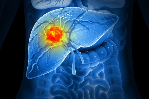
A multicenter clinical trial compared the efficacy of 2-dimensional (2D) and 3-dimensional (3D) contrast-enhanced (CE) ultrasonography (US) evaluations with the current clinical standard of CE magnetic resonance imaging (MRI) or computed tomography (CT) in detecting residual viable hepatocellular carcinoma following transarterial chemoembolization (TACE).
Although research exists on the diagnostic performance of 2D and 3D CE US compared with CE MRI or CT, investigators determined that a multicenter trial was necessary.
Eligible patients over 21 years of age underwent baseline 2D and 3D CE US before receiving TACE and 1 to 2 weeks and/or 4 to 6 weeks after TACE. The patients also received CE MRI or CT 4 to 6 weeks after TACE.
Three fellowship-trained radiologists evaluated CE US and CE MRI or CT for residual viable tumors and compared the results with reference standards of pathology (18%), angiography following re-treatment after discovery of residual disease at 1- to 2-month follow-up imaging (31%), CE MRI or CT at 4 to 8 months follow-up (42%), or CE MRI or CT if clinically decompensated with a viability higher than 50% (9%).
Of the 132 eligible patients, 87 were male and 45 were female. The average patient age was 64 years.
Four to 6 weeks after patients received TACE, the sensitivity of 2D CE US was 91% (95% Cl, 84-95), higher than CE MRI or CT at 68% (95% Cl, 58-76; P<.001). Following the same time frame, 3D CE US sensitivity was 89% (95% Cl, 81-94), also higher than CE MRI or CT.
However, at 4 to 6 weeks, the specificity of CE MRI or CT at 85% (95% Cl, 76-91) was higher than 2D CE US at 70% (95% Cl, 56-80) and 3D CE US at 67% (95% Cl, 53-78).
For 2D CE US, the researchers observed no differences in diagnostic criteria between 1 to 2 weeks and 4 to 6 weeks.


 © 2025 Mashup Media, LLC, a Formedics Property. All Rights Reserved.