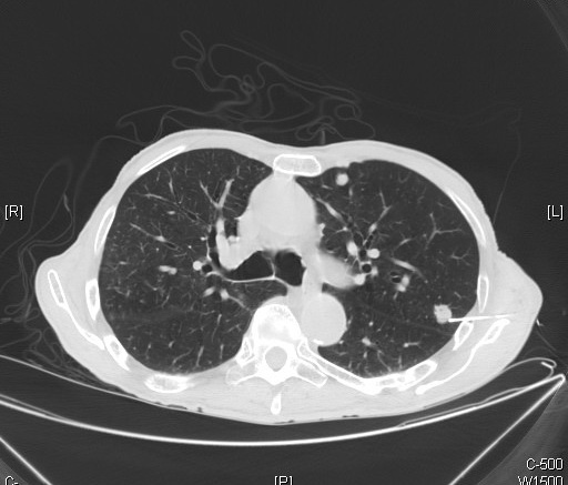
A recent study sought to find the local relapse sites of a group of prostate cancer patients to focus on individualized management conclusions for radiation therapy. By using 18F-PSMA-1007-PET/CT, relapsed tumors can easily be found through hybrid imaging. Researchers aimed to further the use of PSMA to evaluate and map tumor relapse.
While 68Ga-PSMA-11 has been used in the past to find relapse sites and yields a high detection rate, but the tracer is quickly excreted by the urinary tract, making detection challenging. 18F-PSMA-1007-PET/CT has a low renal excretion tract which reduces the risk of inconclusive results as opposed to 68Ga-PSMA-11.
The study included 135 prostate cancer patients who had received primary treatment and underwent 18F-PSMA-1007-PET/CT due to detection of a local relapse through biochemical relapse. The patients’ imaging data was analyzed and reassessed regarding the relapse locations. In the study, 69.6% of patients had undergone prostatectomies, and 30.4% of patients underwent radiation therapy.
The most common finding made with PET imaging was a unifocal relapse in 72.6% of patients. There was also a higher rate of ipsilateral cases in the relapsed tumors. The tumors relapsed most commonly in the posterior region after surgery and in the peripheral zone after radiation therapy.
The study concluded that 18F-PSMA-1007-PET/CT is a viable option for finding the localization and allocation of relapse in prostate cancer patients.
Reference


 © 2025 Mashup Media, LLC, a Formedics Property. All Rights Reserved.