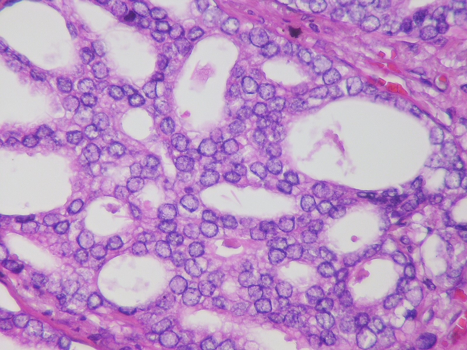
Researchers analyzed a case of a 66-year-old patient who was diagnosed with prostate cancer infiltrating the rectum along with solitary rectal metastases that were staged using 18F-PSMA-1007 PET/CT.
The whole-body scan was able to detect multiple foci of abnormal tracer uptake in the pelvis and abdomen. The PET/CT and contrast-enhanced computed tomography images showed an increased uptake of radiotracer in primary tumor in residual prostate gland infiltrating the neck of urinary bladder, rectum, levator ani, and obturator muscles with pelvic lymphadenopathy.
The patient received six cycles of docetaxel-based chemotherapy with androgen deprivation treatment. After the six cycles of chemotherapy, 18F-PSMA-1007 PET/CT showed persistent radiotracer uptake in the primary and pre-existing metastatic sites in the pelvis.
The rectum is one of the least commonly involved sites of prostate cancer through direct invasion or metastasis, despite its close proximity to the prostate gland. The majority of prostate cancers with rectal involvement present with metastatic disease, but rectal involvement by prostate cancer does not always predict a worse outcome in hormone treatment-naïve patients.
Overall, 18F-PSMA-1007 PET/CT is an accurate staging process for detecting rectal metastases in patients with prostate cancer by showing radiotracer uptake in tumors.
Reference
Isolated Rectal Metastases from Locally Advanced Carcinoma Prostate Detected by 18F-PSMA-1007 PET/CT


 © 2025 Mashup Media, LLC, a Formedics Property. All Rights Reserved.